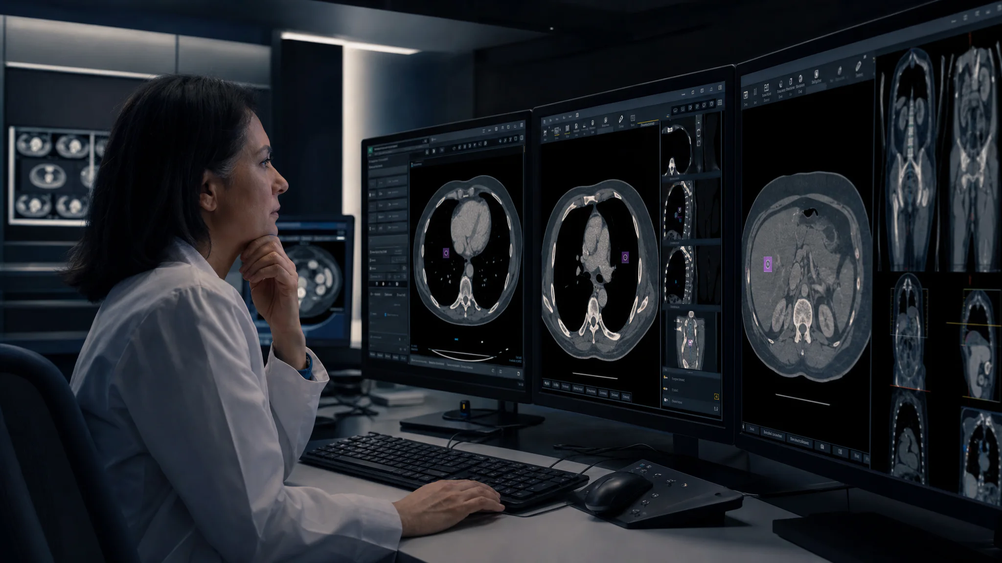
Radiology AI succeeds or fails in the space between the algorithm and the radiologist. A model can be accurate in a study and still fail in practice if images do not route reliably, results open in a separate viewer, patient context is mismatched, or nobody notices performance drift.
This guide is for radiology administrators, imaging informatics teams, PACS managers, and clinical leaders planning a first deployment or cleaning up a collection of disconnected AI tools.
Quick answer: require standards-based interfaces, map the complete order-to-report workflow, validate on local studies, keep results inside the normal reading environment, capture radiologist corrections, and monitor performance after every material change.
The reference architecture in plain language
A dependable post-acquisition workflow looks like this:
The order-to-report loop, with AI inside it
01
Order
The EHR or RIS creates and identifies the imaging order
02
Acquire
The modality sends DICOM objects to PACS or a VNA
03
Route
An orchestrator selects eligible studies and copies them to the right AI service
04
Return
The AI result comes back in a format the viewer and reporting system can use
05
Read
The radiologist accepts, edits, or rejects inside the normal workflow
06
Monitor
Outcomes, corrections, errors, and timing feed monitoring
 A standards-based radiology AI architecture from modality through PACS and back to the radiologist
RSNA's Imaging AI in Practice demonstration used DICOM, HL7, FHIR, IHE profiles, standardized event logs, and common data elements to move orders, images, AI outputs, and reports through a simulated clinical workflow. Its practical recommendation is direct: interoperability standards should appear in RFPs and contracts. See the RSNA workflow demonstration.
Map the workflow before selecting a model
Document one representative study from order to signed report. Include systems, owners, identifiers, statuses, and timing. Then mark the exact moments where AI should act.
Questions to answer:
- Which procedure codes or DICOM attributes make a study eligible?
- Does AI run before worklist triage, before interpretation, during reporting, or after sign-off?
- What happens when a series is incomplete or arrives late?
- Where does the radiologist see the output?
- Can the result prepopulate a report, and can it be changed?
- How are critical results communicated without creating duplicate alerts?
- What is the fallback if the AI service is slow or unavailable?
- Which system is the source of truth for the final clinical record?
This map prevents a common mistake: buying an algorithm for a finding without deciding how that finding changes the worklist, viewer, report, or patient pathway.
The interfaces your RFP should name
| Standard or interface | What it usually carries | What to verify |
|---|---|---|
| DICOM C-STORE and DICOMweb | Images and derived objects | Supported objects, transfer syntax, metadata preservation, retry behavior |
| DICOM SR, SEG, and secondary capture | Structured findings, segmentations, visual results | Whether your PACS displays and preserves them correctly |
| HL7 v2 | Orders, statuses, reports, patient updates | Trigger events, identifier mapping, acknowledgements, correction flow |
| FHIR | Modern clinical data and workflow APIs | Exact resources, profiles, authorization, and write-back capability |
| IHE AI Workflow for Imaging | Task orchestration between requester, manager, and performer | Which actors and transactions each vendor supports |
| SSO and audit interfaces | Identity, access, and traceability | Role mapping, break-glass behavior, immutable audit data |
Do not accept "supports DICOM" as a complete answer. Ask which services, information objects, tags, and error conditions were tested with your PACS version.
Decide how results return to the radiologist
The result format changes both usability and safety.
Worklist priority
Useful for time-sensitive findings, but it can create alert fatigue and unintended queue behavior. Define maximum delay, failure handling, and what happens when AI is negative or unavailable.
Viewer overlay or segmentation
Useful when the radiologist needs spatial context. Confirm that overlays align after image transformations and remain clearly distinguishable from source images.
Structured measurement or report field
Useful for repeatable measurements and reporting efficiency. The radiologist must be able to accept, edit, or reject the value, and the final report must preserve who made the decision.
Separate application
Sometimes unavoidable during a pilot, but it adds context switching and makes adoption harder. Treat it as a temporary constraint with a defined integration plan.
A multi-society statement from ACR, CAR, ESR, RANZCR, and RSNA warns that extra applications add workload, local performance may not match vendor claims, and post-market monitoring is necessary. It also recommends local validation before implementation and repeat assessment after relevant software or equipment changes. Read the full practical considerations statement.
Local validation: define pass and fail before the pilot
Select a retrospective sample that reflects your scanners, protocols, patient population, sites, and disease prevalence. Include known difficult and low-quality cases. Lock the acceptance criteria before seeing the results.
Measure more than sensitivity and specificity
- Failed and excluded study rate
- Time from image completion to available result
- Wrong study, series, or patient association events
- Performance by site, scanner, protocol, and patient subgroup
- Radiologist acceptance, modification, and rejection rates
- Effect on report turnaround and worklist behavior
- False alerts per shift or per reading session
For a prospective silent phase, run the tool without showing results to radiologists. This exposes routing, latency, and data-quality problems without influencing care. Move to a visible pilot only after the silent acceptance criteria pass.
Monitoring is part of the clinical system
 A radiology quality team reviewing local AI concordance and drift
The 2026 ACR-SIIM imaging AI practice parameter and Assess-AI framework emphasize selection, local evaluation, monitoring, and continuous quality improvement. ACR describes concordance tracking between AI outputs and report-derived labels, with local review of discordant cases. See the ACR announcement and framework overview.
Your minimum monitoring view should show:
- Eligible, processed, failed, and timed-out studies
- Result latency by site and modality
- Concordance or clinically reviewed quality signal
- Correction and override patterns
- Performance by scanner, protocol, site, and subgroup where feasible
- Software, model, and configuration version
- Incidents, investigations, and corrective actions
Set thresholds that create a review, a restricted-use state, or a pause. A dashboard without an owner and response rule is only decoration.
The hidden integration risks
Patient and study matching
Use stable identifiers and validate accession, study, series, and patient context at every handoff. Quarantine ambiguous matches instead of guessing.
Incomplete data
Late series, protocol changes, localizer images, and outside studies can trigger incorrect routing. Define readiness rules for each use case.
Automation bias
Train radiologists on the intended use, known limitations, and appropriate disagreement. Preserve an easy way to reject or correct results.
Updates outside your control
Contract for advance notice of model, threshold, hosting, interface, and intended-use changes. Decide which changes require interface regression testing, local revalidation, or governance approval.
Cybersecurity and downtime
Document network paths, data destinations, authentication, encryption, logging, patching, backup, and recovery. The reading workflow must remain safe when AI is unavailable.
A practical 90-day implementation plan
Weeks 1–3
Workflow and interfaces
Choose one use case and one clinical owner. Map the current workflow, define intended use, inventory systems, confirm regulatory status, and obtain interface specifications from every vendor.
Weeks 4–6
Technical integration and silent run
Build routing, result return, logging, and failure handling in a non-production environment. Validate identifiers and object compatibility. Begin a prospective silent run.
Weeks 7–9
Local clinical validation
Review the predefined sample, analyze subgroups and technical failures, set thresholds, and complete training. Resolve workflow defects before showing results in care.
Weeks 10–12
Limited launch
Release to a small trained group. Monitor daily at first. Review overrides and incidents with the clinical owner. Expand only after the operational and clinical criteria remain stable.
Copy this radiology AI RFP checklist
- State the cleared or authorized intended use and current model version.
- List every supported DICOM service and result object.
- List HL7, FHIR, IHE, SSO, and audit capabilities.
- Demonstrate the complete workflow in our PACS, RIS, viewer, and reporting stack.
- Explain study eligibility, incomplete-series handling, retries, and downtime behavior.
- Show how radiologists accept, edit, reject, and report AI results.
- Provide local validation support and machine-readable result export.
- Define monitoring metrics, update notices, incident response, and version rollback.
- Identify all data locations, subprocessors, retention periods, and security controls.
- Quote interface, implementation, support, and three-year operating costs.
The decision
The best radiology AI integration is almost invisible during a normal read and highly visible when quality teams need evidence. Standards reduce custom interface debt, but local workflow design, validation, and monitoring make the system clinically usable.
If your PACS, RIS, vendor algorithms, and reporting environment do not connect cleanly, the work is not another model comparison. It is an integration architecture problem. That is where an imaging-focused engineering partner can turn a promising algorithm into a dependable clinical workflow.